 Cell Technology Co., Ltd.")

News
News Center
Stem cells—The new force restoring the gift of sight
2022-06-06
If God granted you three days of sight, where would you choose to fix your gaze the most? In her famous book *Three Days to See*, American author Helen Keller answered this question beautifully: "I would surely linger over the things I hold dear, so that in the darkness ahead, I could recall them one by one." What a poignant, almost tragic, and profoundly melancholic choice it is—yet today, thanks to advances in biotechnology and stem-cell research, we stand on the brink of a new chapter in treating eye diseases.
Modern treatments for various eye conditions are advancing rapidly. In recent years, ophthalmologists have increasingly focused their attention on stem-cell research. Today, stem cells have already made significant strides in the treatment of many types of eye disorders.
1. Myopia
With the advancement of technology, televisions, smartphones, and tablets have become ubiquitous, enriching our perspectives while significantly cutting into people's time spent on outdoor activities. According to data released by the National Health Commission in July 2021, the overall myopia rate among children and adolescents in China reached 52.7% in 2020, an increase of 2.5% compared to 2019.
The age of onset for myopia is getting younger, and the number of people with high myopia is also on the rise. There is an urgent need for effective treatments that can halt the progression of myopia during childhood and help reduce its overall prevalence.
Mesenchymal stem cells have been successfully applied in various clinical settings for connective tissue regeneration and reconstruction, and their unique properties make them an ideal cell candidate for scleral reinforcement. [1] Since the pathological mechanisms underlying myopia progression (as shown in Figure 1) align with the characteristics of stem cells, sub-scleral injection of mesenchymal stem cells can help strengthen the already fragile sclera in myopic patients. The transplanted cells are expected to differentiate into fibroblasts, promoting the production of extracellular matrix that reinforces the sclera and inhibits eye elongation—thereby preventing or halting the progression of myopia (as illustrated in Figure 2).
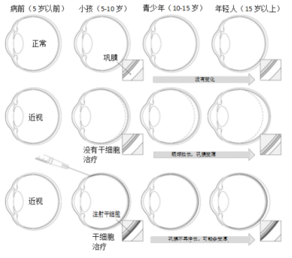
Figure 1: A schematic illustration of the progression of severe myopia, along with the potential role of stem cell therapy in halting disease advancement. Note how, over time, the axial length of the eye elongates, the sclera thins, and the promising outlook for preventing further disease progression when treatment is initiated early.
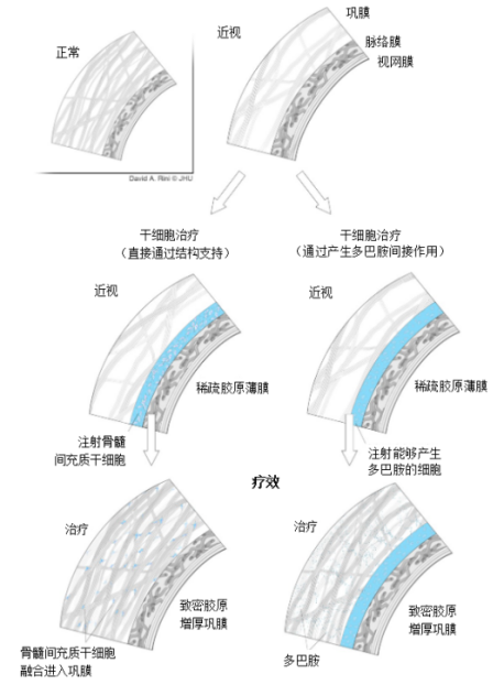
Figure 2: Stem cell-based therapies could potentially harness mechanisms that prevent myopia progression. These approaches include combining injected mesenchymal stem cells with the scleral structure for direct mechanical support, as well as indirectly stimulating scleral tissue by promoting dopamine production, thereby inhibiting eye elongation.
II. Retinitis Pigmentosa
Retinitis pigmentosa (RP) is a group of inherited eye diseases caused by damage and eventual death of retinal cells, leading to progressive vision loss. Since the optic nerve and retinal ganglion cells lack the ability to regenerate, irreversible damage occurs once these cells have been irreversibly lost through apoptosis.
The primary mechanism by which mesenchymal stem cells exert their effects in degenerative diseases is through the secretion of growth factors that modulate the paracrine microenvironment. These cells serve as a source of trophic factors, promoting cell survival and activating intrinsic repair mechanisms. [2] Because MSCs do not express the major histocompatibility complex (MHC) on their cell surfaces, this advantage enables autologous or allogeneic use without the risk of rejection. As a result, patients do not require any immunosuppressive therapy after transplantation to prevent rejection reactions. [3-4] 。
A Phase 3 clinical trial published on the Research Gate website was conducted by experienced surgeons using Limoli’s retinal repair technique (LRRT), performed under local anesthesia. Limoli and colleagues provided a detailed description of this approach. [5-7] , the stem cell suspension was injected into the fat tissue located between the transplanted choroid and sclera. During the procedure, each eye received 5 million UC-MSCs, and the patients were followed up for six months (as shown in Figures 3 and 4). [8] 。
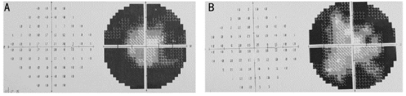
Figure 3: Visual field measurements of the patient before treatment (A) and 6 months post-treatment (B), showing a reduction in peripheral visual field defects during the study period.

Figure 4: Pre-treatment (A) and 6-month post-treatment multifocal electroretinogram (mfERG) recordings (B)—color maps and 3D visualizations—demonstrate improved mfERG responses, particularly in the central ring area.
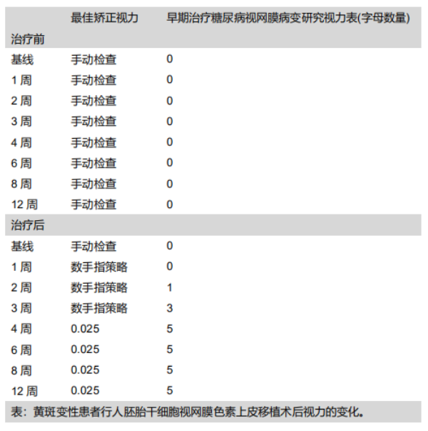
In 2010, the U.S.-based company ACT (Advanced Cell Technology) received FDA approval to use retinal pigment epithelium (RPE) cells derived from embryonic stem cells for treating two degenerative eye diseases: dry age-related macular degeneration (Dry AMD) and Stargardt disease. This marked the second clinical trial approved by the FDA involving human embryonic stem (hES) cell-derived therapies.
In 2012, the research institution published an article titled "Embryonic stem cell trials for macular degeneration: a preliminary report" in The Lancet magazine. [9] U.S. researchers have used human embryonic stem cells (hESCs) to treat two patients suffering from distinct degenerative eye diseases. Initial data suggest that hESC therapy is not only safe but also shows promising therapeutic potential. The team first induced the hESCs to differentiate into early bone cells and neural tissue cells, which subsequently matured into retinal pigment epithelial cells—achieving a purity level exceeding 99%.
Researchers injected approximately 50,000 isolated retinal pigment epithelial (RPE) cells beneath the retinas of two patients: a senior woman who developed dry age-related macular degeneration in her 70s, and a middle-aged woman suffering from Stargardt disease, both of whom had already become blind. Four months later, the researchers observed that the RPE layer had been completely replaced by the injected RPE cells. The data confirmed that the transplanted cells had successfully survived and significantly improved the patients' vision. (See table.)
3. Macular Degeneration
Macular degeneration is typically a natural consequence of age-related deterioration; as we grow older, the retinal tissue breaks down and thins, leading to a decline in macular function. Macular degeneration is categorized into two types: age-related macular degeneration (ARMD) and juvenile macular degeneration. In recent decades, the incidence of age-related macular degeneration has been steadily rising, making it one of the leading causes of blindness among people over 60. [10-11] 。
Macular degeneration is characterized by the loss of retinal pigment epithelial (RPE) cells, which subsequently leads to degenerative changes in photoreceptor cells such as cones and rods. This progressive deterioration gradually erodes a patient’s visual acuity, making it increasingly difficult to read, view newspapers, or even drive. Unfortunately, current clinical treatments are unable to address the root cause of the disease, and some patients show poor responses to these therapies. As a result, researchers are now exploring innovative approaches to treat macular degeneration—specifically, using stem cell therapy to replace the dying RPE cells in patients’ eyes, potentially preserving vision before irreversible damage occurs.
RPE can be derived from human embryonic stem cells (hESCs) or human induced pluripotent stem cells (iPSCs). [12-14] Before photoreceptors suffer irreversible damage, replenishing lost or degenerated RPE cells with RPE cells differentiated from stem cells can restore retinal function. The differentiated cells exhibit a morphology similar to native RPE cells, express RPE-specific markers, and retain the ability to phagocytose photoreceptor outer segments. [15] 。
An article published in *Nature Biotechnology* [16] Researchers have developed a retinal pigment epithelium (RPE) patch derived from stem cells, which holds promise as a treatment for sudden, severe vision loss caused by macular degeneration. The team used specially designed microsurgical tools to implant the patch into the subretinal space of one eye in two patients suffering from advanced exudative macular degeneration. Using advanced bio-microscopy and optical coherence tomography, the RPE patches were successfully integrated and remained viable at 12 months post-surgery. Notably, both patients experienced significant improvements in visual acuity—gaining 29 and 21 letters, respectively—demonstrating the potential of this approach. This clinical study provides critical evidence supporting the feasibility and safety of human embryonic stem cell-derived RPE patch transplantation as a therapeutic option for age-related macular degeneration.
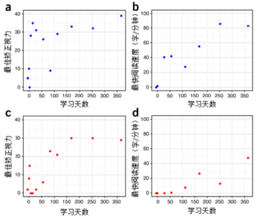
Figure 5: Best Corrected Visual Acuity and Reading Speed for Cases 1 and 2
(a) The patient's best-corrected vision has remained stable for over 12 months.
(b) Patient 1’s reading speed exceeds 12 months.
(c) Patient 2 achieved best-corrected visual acuity lasting over 12 months.
(d) Patient 2’s reading speed has improved beyond the 12-month mark.
4. Glaucoma
Glaucoma is a neurodegenerative disease characterized pathologically by optic nerve atrophy and visual field defects, leading to impaired vision and, in severe cases, even blindness. The underlying mechanisms involve abnormally high intraocular pressure and insufficient blood supply to the optic nerve.
Currently, treatment options involve lowering intraocular pressure through medication or surgery, along with neuroprotective therapies. While these approaches can provide some protective benefits, they are unable to fundamentally prevent damage to the optic nerve.
Researchers have found that treating animal models of optic nerve injury with mesenchymal stem cells yields significant therapeutic effects. The mechanism involves homing mesenchymal stem cells, which secrete cytokines via paracrine signaling to help repair the damaged nerve cells. [17] 。
In 2013, the journal *Stem Cells* published an article titled "Transplantation of Mesenchymal Stem Cells Promotes Tissue Regeneration in a Glaucoma Model Through Laser-Induced Paracrine Factor Secretion and Progenitor Cell Recruitment." [18] 。
Researchers from Canada transplanted mesenchymal stem cells into a glaucoma model, demonstrating that laser-induced secretion of paracrine factors and enhanced progenitor cell proliferation can promote tissue regeneration in glaucoma. The experiments revealed that mesenchymal stem cells derived from bone marrow stimulate the regeneration of trabecular meshwork cells, with intracameral injection proving significantly more effective at reducing intraocular pressure (IOP) compared to hematopoietic cells (p < 0.001), as shown in Figure 6. Additionally, mesenchymal stem cells and the factors they secrete reawaken the pool of progenitor cells within the ciliary body, thereby boosting cellular proliferation, as illustrated in Figure 7.
Laser-induced tissue remodeling directs mesenchymal stem cells precisely to the damaged area and also leads to a modest increase in ocular progenitor cells. This study demonstrates that mesenchymal stem cells and their secreted factors serve as critical mediators for tissue repair in open-angle glaucoma, specifically through the recruitment of local neural progenitor cells.
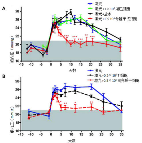
Figure 6: MSC (mesenchymal stem cells) induction experiment shows that intraocular pressure in experimental glaucoma rapidly returned to normal.
(A): Intraocular injection of 1×10⁶ bone marrow mononuclear cells (red), 1×10⁶ lymphocytes (black), and saline solution (green); no additional treatment was administered (blue). The gray area indicates the normal range for intraocular pressure. Intraocular pressure values represent the mean ± SEM from four experiments, with 12 animals evaluated per group.
(B): 0.5 × 10^6 T cells (in black) or 0.5 × 10^6 mesenchymal stem cells (in red) were injected intraocularly after laser irradiation, and the results were evaluated as described above. The average values ± SEM from three experiments, each involving 9 animals per group, were calculated.
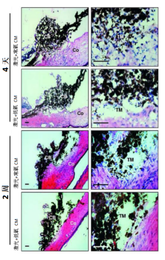
Figure 7: Hematoxylin and Eosin (H&E) staining of the anterior segment of rat eyes immediately after treatment with laser irradiation of normoxic or hypoxic MSC-CM (conditioned medium). Evaluations were conducted on days 4 and 14 post-MSC-CM injection.
5. Cataracts
Cataracts occur when the lens inside the eye becomes cloudy, changing from clear to opaque and blocking light from entering the eye, thereby impairing vision. In the early stages, mild cloudiness or a small affected area may not impact sight—but over time, the condition progressively worsens, eventually leading to significant vision loss or even blindness. Unfortunately, vision impairment caused by cataracts cannot be corrected with eyeglasses alone. According to the World Health Organization, approximately 20 million people worldwide are blind due to cataracts, making it the leading cause of blindness globally.
Traditional treatment methods typically involve using ultrasound to soften and break up the lens, then flushing it out of the eye. Afterward, doctors implant an artificial intraocular lens into the patient’s eye. However, this approach can sometimes lead to severe postoperative inflammation and complications—potentially even resulting in irreversible blindness. Moreover, artificial lenses are not only expensive but also lack the delicate, precise physiological regulation capabilities of the natural lens. As a result, patients often experience glare after surgery, which can significantly impair visual quality. [19] These inherent defects in artificial lenses are driving researchers to explore new treatments for cataracts.
Professor Yi-Zhi Liu, leading a team from the Zhongshan University Eye Center, has discovered that the human eye’s lens contains endogenous stem cells. Using these unique stem cells, the researchers successfully regenerated a transparent lens in situ—marking the first-ever achievement of functional, physiologically active tissue regeneration in humans. This groundbreaking approach has now been applied clinically to treat congenital cataracts. The findings were published in March 2016 in the prestigious journal *Nature*. [20] In the study, 12 infants under the age of 2 who were born with congenital cataracts underwent this entirely new surgical procedure. Post-surgery, functional lenses successfully regenerated, and the recurrence rate was reduced by more than 20-fold. This clinical trial not only confirmed the safety and effectiveness of the innovative technique in treating congenital cataracts but also offers a groundbreaking strategy for cataract treatment, while simultaneously opening up new avenues for tissue regeneration and the clinical application of stem cells.
In addition to this, treatments involving the transplantation of limbal stem cells, mucosal epithelial cells from the oral cavity, and corneal epithelial stem cells are being used to repair corneal injuries. Meanwhile, research is actively underway on using embryonic stem cell-derived retinal pigment epithelial (ES-RPE) cells, as well as hematopoietic and mesenchymal stem cell transplants, to address certain autoimmune-related eye disorders.
Scientists and researchers around the world have never stopped pursuing the path to light, confident that in the near future, stem cells will unlock cures for more eye diseases—and bring hope and vision back to those who suffer from them.
References:
[1] MIROSLAW JANOWSKI, Jeff W M Bulte. Concise Review: Using Stem Cells to Prevent the Progression of Myopia—A Concept. STEM CELLS 2015;33:2104–2113 www.StemCells.com.
[2] Ng TK, Fortino VR, Pelaez D, et al. Progress in mesenchymal stem cell therapy for neural and retinal diseases. World J Stem Cells 2014; 6: 111–119.
[3] Nauta AJ and Fibbe WE. Immunomodulatory properties of mesenchymal stromal cells. Blood 2007; 110: 3499–3506.
[4] Kyurkchiev D, Bochev I, Ivanova-Todorova E, et al. Secretion of immunoregulatory cytokines by mesenchymal stem cells. World J Stem Cells 2014; 6(5): 552–570.
[5] Limoli PG, Limoli C, Vingolo EM, Scalinci SZ, Nebbioso M. Cell surgery and growth factors in dry age-related macular degeneration: visual prognosis and morphological study. Oncotarget 2016;7(30):46913-46923.
[6] Limoli PG, Vingolo EM, Morales MU, Nebbioso M, Limoli C. Preliminary study on electrophysiological changes following cellular autograft in age-related macular degeneration. Medicine (Baltimore) 2014;93(29):e355.
[7] Limoli PG, Vingolo EM, Limoli C, Nebbioso M. Stem cell surgery and growth factors in patients with retinitis pigmentosa: a pilot study following a literature review. Biomedicines 2019;7(4):E94.
[8] Neslihan Sinim Kahraman, Ayse Oner. Umbilical Cord-Derived Mesenchymal Stem Cell Implantation in Retinitis Pigmentosa: 6-Month Follow-Up Results of a Phase 3 Trial. DOI:10.18240/ijo.2020.09.14.
[9] Schwartz SD et al. (2012): Embryonic stem cell trials for macular degeneration: a preliminary report, The Lancet, 379(9817):713-720.
[10] Friedman DS, O’Colmain BJ, Munoz B, Tomany SC, McCarty C, de Jong PT, et al. Prevalence of age-related macular degeneration in the United States. Arch Ophthalmol 2004; 122(4): 564-72.
[11] Klein R, Klein BE, Lee KE, Cruickshanks KJ, Gangnon RE. Changes in visual acuity in a population over a 15-year period: the Beaver Dam Eye Study. Am J Ophthalmol 2006; 142(4): 539-49.
[12] Carr AJ, Vugler AA, Hikita ST, et al. Protective effects of human iPS-derived retinal pigment epithelium cell transplantation in the retinal dystrophic rat. PLoS ONE 2009;4:e8152.
[13] Cho MS, Kim SJ, Ku SY, et al. Generation of retinal pigment epithelial cells from human embryonic stem cell-derived spherical neural masses. Stem Cell Research 2012;9:101–9.
[14] Buchholz DE, Hikita ST, Rowland TJ, et al. Derivation of functional retinal pigmented epithelium from induced pluripotent stem cells. Stem Cells 2009;27:2427–34.
[15] Jakub Hanus, Fangkun Zhao, Shusheng Wang. Current therapeutic advances in atrophic age-related macular degeneration. Br J Ophthalmol 2016;100:122–127. doi:10.1136/bjophthalmol-2015-306972.
[16] Phase 1 clinical study of an embryonic stem cell–derived retinal pigment epithelium patch for age-related macular degeneration. Received November 6, 2016; accepted February 28, 2018; published online March 19, 2018; doi:10.1038/nbt.4114
[17] Chung S, Rho S, Kim G, et al. Human umbilical cord blood mononuclear cells and chorionic plate-derived mesenchymal stem cells promote axon survival in a rat model of optic nerve crush injury[J]. International Journal of Molecular Medicine, 2016, 37(5): 1170-1180.
[18] Mesenchymal Stem Cell Transplantation Promotes Tissue Regeneration in a Glaucoma Model by Inducing Laser-Driven Paracrine Factor Secretion and Recruiting Progenitor Cells. Renaud Manuguerra-Gagné, Patrick R. Boulos.
[19] Liu Yizhi, Dong Xia. Stem Cells and Lens Regeneration. Chinese Journal of Cell and Stem Cell (Electronic Edition), February 2014, Vol. 4, No. 1.
[20] Haotian Lin, Hong Ouyang. Lens Regeneration Using Endogenous Stem Cells with Restoration of Visual Function. Nature, Volume 531, pages 323–328 (2016). DOI: 10.1038/nature17181.
Related News

Here is the title—h1 placeholder text


Copyright © Jiuzhitang Maker (Beijing) Cell Technology Co., Ltd.
Powered by: 300.cn SEO | Privacy Policy